Cell Culture Monitoring
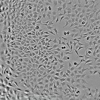
Constant cell culture monitoring serves to ensure
- Cell health
- Growth rate
- Morphology
- Stability of the cell culture system
Automize cell imaging
Determine cell confluence, cell cycle phases, metabolic activity and contamination status automatically with our live cell imager. Identify the ideal cell confluence of every cell line as the perfect starting point for your experiments without error-prone subjective interpretation. Use our automatic cell alarms to have full control and be able to intervene immediately.
Minimize downtime, increase reproducibility and improve the quality of your cell cultures without any hands on work. And the best thing: You can analyze your cells directly in the incubator! Prevent fluctuations of conditions (e.g. temperature, CO₂-supply and humidity) and minimize contamination risk at the same time!
Monitor 24 wells in paralell
With 24 miniature microscopes our live cell imager monitors up to 24 cell cultures in paralell. You can scan all 24 wells in less than 30 sec and analyze them within 5 min. Raise the validity of your experiments, compare multiple wells simultanously and improve reproducibility.
Analyze your cells remotely
Get real-time data 24/7 even for long term periods. Don’t miss any change in cell morphology and visualize your cells while they are dividing, moving or dying through zooming to single cell level. Use time laps videos for your documentation.
Monitor and analyze your cells remotely. At any time and any place you want!
Scratch Assays
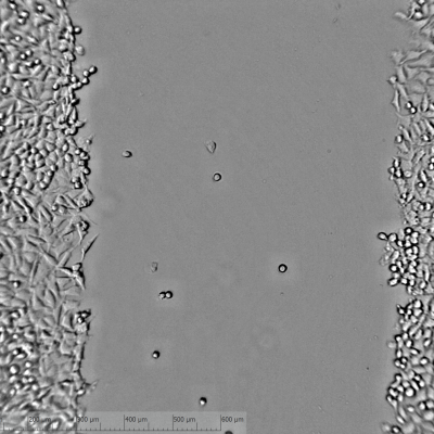
Scratch assay is a standard method to study cell migration in vitro. A small “wound” or gap is created in a cell layer to observe how the cells migrate into the free space to close the gap and to determine the gap closure time. This is an easy way to test the effects of drugs, genetic changes or other factors on cell migration and an important method for wound healing and cancer research.
Visualize the full process
Our live cell imager records your migrating cells every 4 min (or longer intervalls) and enables a full enclosure of the migration process on single cell level. Don’t miss anything of the wound healing process and calculate the speed of wound healing by analyzing the increase of confluence in the gap area.
Analyze up to 24 migration assays simultanously
Analyze up to 24 different migration assays under exact identical conditions and optimize comparability such as reproducibility of your results.
Cytotoxicity Assays
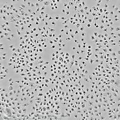
Cytotoxicity assays are important tools to determine the effect of drug candidates on cells. A central aspect of these assays is the observation of morphological changes in the cells, which indicate cell damage/death and are often early indicators of toxic effects. By combining morphological observations with cell viability assays, a comprehensive picture of cytotoxic effects can be obtained.
Vizualize drug effects on morphiology
Monitor changes in cell morphology as a result of drug treatment on single cell level, automatically and in long-term. With our live cell imager you can vary image scan intervalls from 4 min to 24 h.
Compare up to 24 different drug conditions
With 24 independent miniature microscopes our live cell imager allows to analyze a set up of 24 different assay conditions in parallel. Compare different drugs or different drug concentrations under identical environment conditions in an automated non-invasive way.
Timelaps Imaging of Spheroids
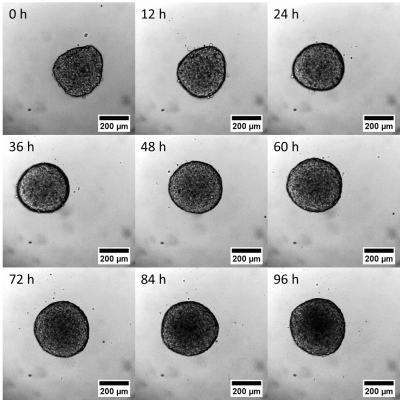
Spheroids - three-dimensional multicellular aggregates - are a very important in vitro model for the investigation of cellular processes. Spheroids mimic the 3D structure of tissues more than 2D cell cultures and create realistic tissue-like environments. They provide better approximation of cell-cell/ cell-matrix interactions and enable more realistic insights into cellular processes for cancer research or the development of new drugs.
Monitor cellular processes in real-time
Our compact and automated live cell imager allows gapless monitoring of spheroids directly within the incubator. Visualize cellular processes with automated timelaps imaging (Interval range: 4 min to 24 h) and test new drug candidates on realistic cell culture models.
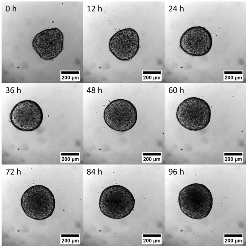
Test multiple drug candidates in one experiment
With 24 independent miniature microscopes our live cell imager allows the analysis of up to 24 spheroid set ups in parallel. Compare different drugs or different drug concentrations in an identical and realistic tissue-environment.
